Tibial Tuberosity Advancement (TTA)
TTA is an orthopedic procedure to repair torn cranial cruciate ligaments in dogs. The function of the cranial cruciate ligament (CrCL) is to stabilize the knee joint, called the stifle in dogs. It limits the tibia from sliding forward in relation to the femur.

Damage to the CrCL is one of the most common injuries in dogs and causes severe lameness. Uncorrected CrCL deficiencies have been associated with meniscal damage and degenerative joint diseases such as osteoarthritis.
The Procedure
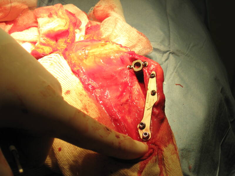
The TTA procedure involves making a cut in the front part of the tibia bone (tibial tuberosity) and advancing this portion of bone forward in order to realign the patellar ligament so that the abnormal sliding movement within the knee joint is eliminated.
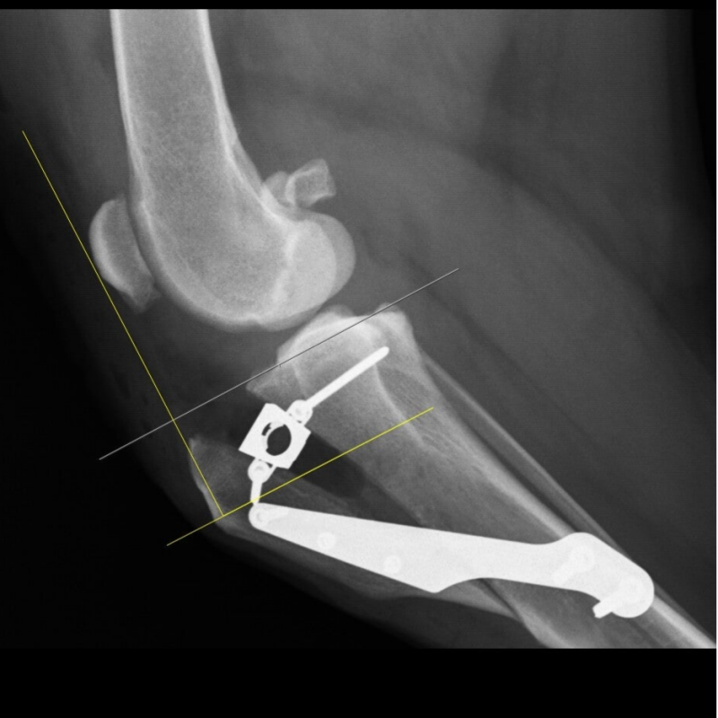
A specialized bone spacer, plate and screws are used to secure the bone in place. Bone graft is collected from the top of the tibia and placed in the gap in the bone to stimulate healing. Recovery restriction time for the TTA requires 12-16 weeks. Generally, dogs are walking near normal by suture removal (10-14 days after surgery).
Call for your evaluation today!
(810) 412-4700
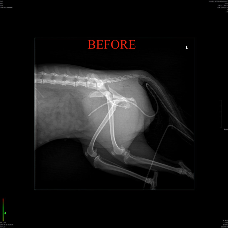
Lateral Suture (MRIT)
The lateral suture technique is one method for repair of a torn cranial cruciate ligament. Two nylon sutures are placed on the lateral side (outside) of the knee (stifle) joint. The sutures are anchored around one of the fabella (small, round bones behind the femur) and passed through a small hole drilled through the tibial tuberosity. This technique is generally considered to be acceptable for dogs under 40-50lbs. Dogs over 50-60lbs generally function much better when an alternative technique, such as tibial tuberosity advancement, is performed.

This shows a radiograph of a dog's stifle joint before the lateral suture has been placed.
Interlocking Nail
The interlocking nail technique can be used when there is a long bone fracture in a bone such as the humerus.
The interlocking nail is a specialized pin that is placed inside the bone and locked into place with bone screws. The screws go through the bone and the nail so that the nail is unable to slide out and the bone fragments cannot rotate around the nail. The nail is left in place for the life of the animal.

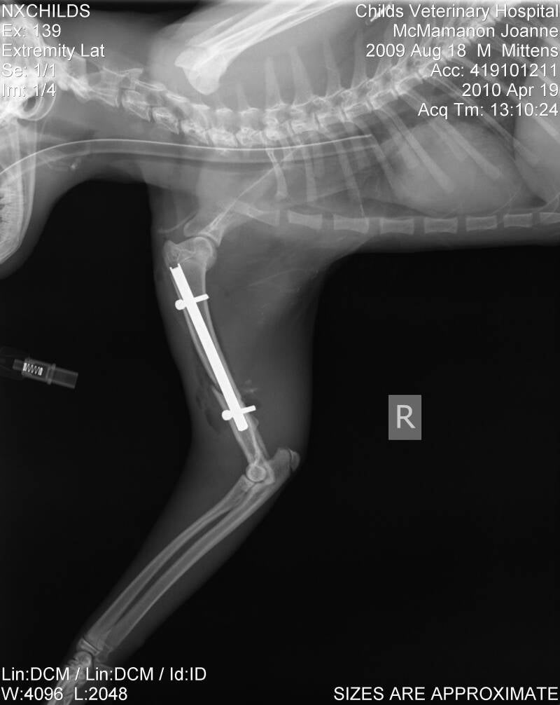
Fracture healing generally requires 8-12 weeks. During this time, activity should be limited so that the repair does not break down. X-rays are taken to assess how the bone is healing. When there is adequate callus formation on the bone, activity is usually allowed unrestricted afterward.
Fracture Repair